Exercise & Bone Health
Bone is living scaffolding that adapts to the loads it is asked to carry — more slowly than muscle, and with a different timetable for men and women. This hub covers the exercise–bone evidence by age, sex, and clinical population: building peak bone mass, what trials show in men, moving after vertebral and hip fractures, and how under-fueling quietly breaks bone in athletes.
What the evidence supports
- Randomized trials show exercise can improve or maintain bone mineral density at the loaded sites — in men, a meta-analysis of RCTs found a small but significant femoral-neck effect (g ≈ 0.21) (Hamilton et al., 2021).
- Expert consensus (UK, 2022) recommends resistance and impact exercise for bone strength, plus strength–balance work to reduce falls and spinal extension for posture — with explicit safety limits after vertebral fracture.
- Bone's response is site-specific: it grows where it is loaded, which is why the femoral neck responds differently from the lumbar spine.
What remains uncertain
- Whether BMD gains from exercise translate into fewer fractures is inferred, not directly demonstrated — fracture outcomes need trials far larger than the field has produced.
- Most high-quality data cluster in postmenopausal women; men, younger athletes, and fracture populations are underrepresented.
- Exercise is adjunct, not treatment: no exercise program has been shown to match pharmacological therapy in established osteoporosis.
Evidence last reviewed: October 6, 2026. Conclusions may change as new research is published.
How Bone Answers Load
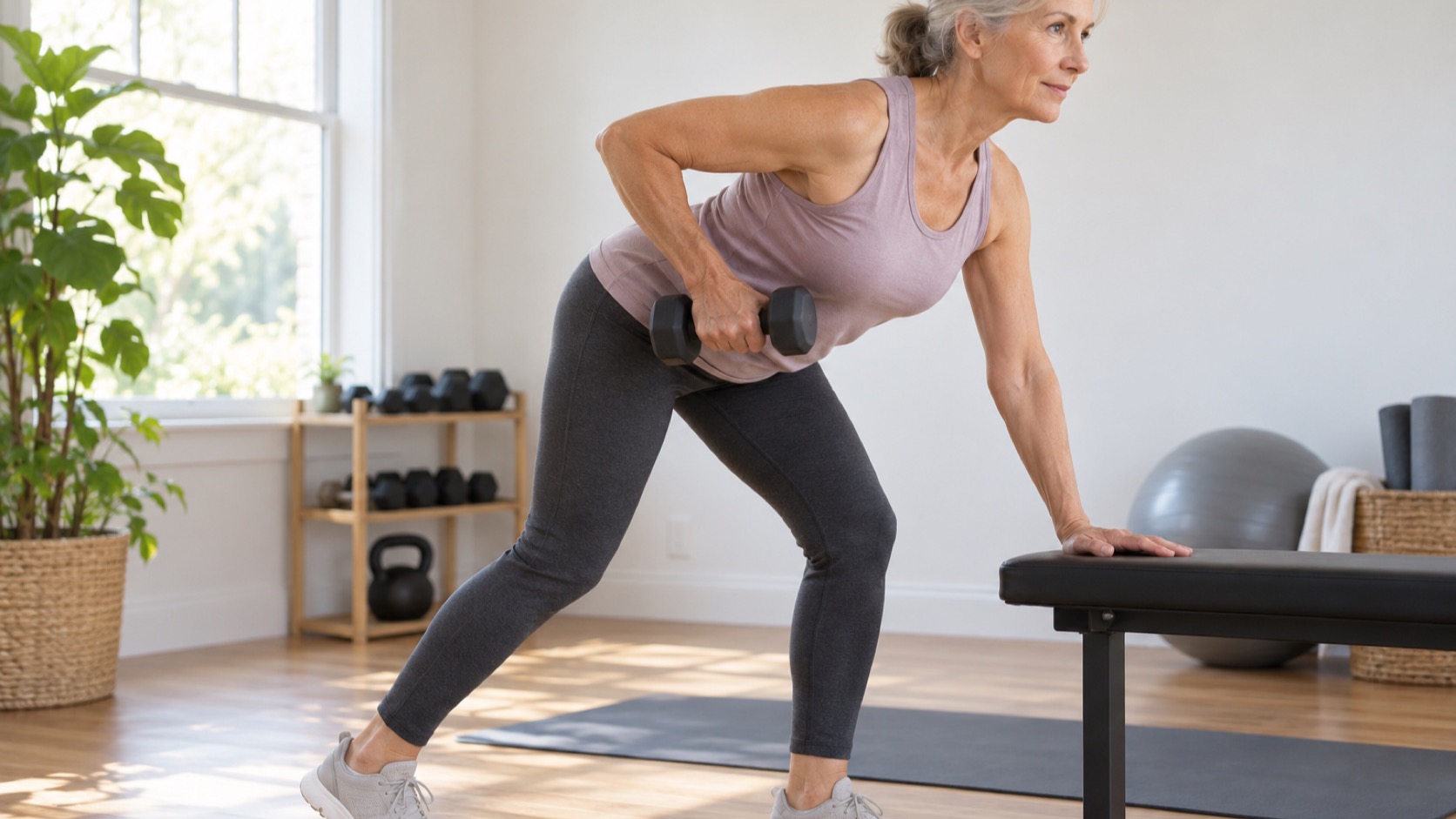
Bone remodels continuously: old tissue is resorbed, new tissue is laid down, and the skeleton is re-tuned to the demands it actually faces. Two forces drive the adaptation — ground reaction force (landing, impact, the jolt of a step) and joint reaction force (muscle pulling on bone through heavy loading). The cells responsible — osteocytes sensing mechanical strain, osteoblasts building, osteoclasts resorbing — respond to strain magnitude and rate, which is why progressive loading matters and why gentle, familiar loads stop stimulating. The general principle and the specific loading programs — heavy lifting, impact work, the LIFTMOR trial — live in the lifting-for-bone subtopic and the Women's Health bone topic; this hub's job is the age, sex, and clinical-population layer on top of that machinery.
The Life Timetable: Peak Mass and Loss
The practical arithmetic of bone health is set early. Most peak bone mass is in place by the late twenties, and the years before it are when the skeleton is most responsive to loading — the "bank account" decade, when impact sports and heavy training deposit bone that later life draws down. After the mid-thirties everyone drifts slowly negative; women face a steep five-to-ten-year loss window around menopause as estrogen withdraws; men lose more slowly, from a higher starting point, and fracture on average a decade later. The distribution matters for expectations: loading in your twenties builds a reserve; loading in your sixties is mostly fighting a slower decline, and exercising after a fragility fracture is rehabilitation, not prevention. That full timetable — and how much of peak mass is modifiable versus genetic — is the peak-bone-mass subtopic's territory.
What the Randomized Trials Show in Men
Men's bone evidence has historically been an afterthought — most exercise–BMD trials recruited postmenopausal women. The best summary of what exists in men is Hamilton et al.'s meta-analysis of randomized trials (>24 weeks; Calcified Tissue International, 2021): a statistically significant benefit at the femoral neck (g = 0.21, 95% CI 0.03–0.40) and no significant effect at the lumbar spine (g = 0.10, −0.07–0.26). The honest reading: real but small, site-specific, and drawn from few trials — the confidence interval for the femoral neck grazes zero, and the authors themselves frame the evidence as limited. What men should actually do with it — loading modes, dose, and how it differs from the women's playbook — is the men's subtopic's territory.
After Vertebral Fracture: The Strong, Steady and Straight Consensus
The most clinically consequential guidance in this territory is the UK consensus statement "Strong, Steady and Straight" (British Journal of Sports Medicine, 2022), built by a multidisciplinary expert group precisely because fear of fracturing again makes people with osteoporosis do less when they usually need to do more, differently. Its shape:
- 🏋️ Strong: resistance and impact exercise to maximise bone strength — scaled to the person, not avoided.
- 🧍 Steady: strength and balance training to reduce falls — the fracture-prevention lever with the strongest direct evidence.
- 🧘 Straight: spinal extension work for posture and possibly lower vertebral-fracture risk.
- ⚠️ Safety rails: avoid end-range loaded spinal flexion (think weighted crunches); after vertebral fracture or multiple low-trauma fractures, keep impact at or below brisk-walking level until clinically cleared.
The exercise progressions, cautions, and return-to-movement details after a vertebral fracture are the spine-safety subtopic's territory; this hub states the frame, that page carries the clinical depth.
Hip Fracture: Where Exercise Is Rehabilitation
A hip fracture is the event the whole skeleton was bracing against, and the exercise question flips from prevention to recovery: rebuilding the ability to walk, stairs, and toilet independence — the functions that decide whether a person goes home. Structured, progressive resistance work during and after rehabilitation is associated with better functional recovery, though trials are heterogeneous and the strongest effects concentrate in supervised, progressed programs rather than generic encouragement to "stay active." The rehabilitation sequence, the evidence, and the return-to-training path are the hip-fracture subtopic's territory.
When Exercise Breaks Bone Instead: Low Energy Availability
The same skeleton that responds to training can be hollowed out by under-fueling. In Relative Energy Deficiency in Sport (REDs — the IOC's 2023 consensus expanded the older Female Athlete Triad into a syndrome affecting men and women), chronically eating too little for the work being done suppresses the hormones bone needs, and BMD falls while training loads continue: stress fractures and, in the worst cases, fragility-grade fractures in twenty-somethings. The warning signs and the diagnostic territory are the low-energy-availability subtopic's territory — the reason it sits in this hub is the caution it puts over everything above: loading builds bone only when the energy and hormonal substrate are there to build with.
The Measurement Problem: BMD as a Stand-In
Nearly every trial in this territory measures bone mineral density, not fractures — and the substitution deserves a moment of scrutiny. BMD is a density snapshot: it says how much mineral sits in a scanned region, not how well the remaining architecture resists a fall, and not whether the person falls at all. Drug trials have shown that BMD gains track fracture reductions reasonably well, which is why BMD is an accepted surrogate — but exercise trials are far smaller than drug trials, run for months rather than years, and produce BMD shifts of a few percent, magnitudes where the fracture translation is an extrapolation rather than a demonstrated result.
The fracture outcome that actually matters has a strong indirect route, though: exercise, especially strength and balance work, reduces falls, and most fractures arrive via a fall. The UK consensus leans on exactly that two-part logic — load the bone, prevent the fall — because each half covers what the other cannot. When someone asks what a training block "bought" their skeleton, the honest answer runs through both channels: a small density shift at the loaded sites, plus a nervous system less likely to deliver the fall that breaks them.
💊 Exercise is not a substitute for osteoporosis treatment
When bone density is already in osteoporosis territory, exercise is an adjunct — never a replacement for evaluation and, where indicated, medication. This is clinician territory: treatment decisions, imaging intervals, and fracture-risk scoring belong to a physician, and the role of exercise in that conversation is to make the pharmacology work on a skeleton that is also being loaded well. Stopping treatment to "try exercise instead" is the failure mode this page exists to prevent.
Questions, Answered Briefly
- Is it ever too late to start loading bone? No — older adults respond to resistance and impact training, just more slowly and from a lower ceiling; the trials in men and in osteoporosis populations show direction, not magnitude.
- Walking — enough? It maintains, mostly; brisk walking is the consensus's impact ceiling after vertebral fracture, but for building, bone wants progressively heavier or faster loading than walking provides.
- Are heavy deadlifts safe with osteoporosis? Sometimes — with diagnosis-appropriate programming and often specialist supervision; the details and the LIFTMOR trial evidence live in the lifting-for-bone and spine-safety subtopics.
- Do men need to think about this? Yes — men fracture too, roughly a decade later on average, and the men's evidence base is thinner than the women's.
The Bottom Line
- Bone adapts to load, site-specifically: what you load is what responds — the femoral neck answers impact and heavy work in men (g ≈ 0.21); the spine's answer is weaker in the trial data.
- The timetable matters: peak-mass years are the deposit window; midlife loading slows loss; post-fracture, exercise is rehabilitation. Same tools, different jobs.
- Consensus exists, and it is more movement, not less: Strong, Steady and Straight — resistance plus impact, strength–balance, spinal extension — with real safety rails after vertebral fracture.
- Exercise is adjunct, never treatment: under-fueling breaks bone (REDs), and established osteoporosis belongs in clinical hands — loading supports therapy, it does not replace it.
Go Deeper: Subtopics
- 🔎 Building peak bone mass — the late-teens-to-late-twenties deposit window: how much of peak mass is trainable, and what the pre-peak training years buy. Read it →
- 🔎 Exercise and bone health in men — the thin male evidence base, the femoral-neck finding, and how men's programming differs from the postmenopausal playbook. Read it →
- 🔎 Spine-safe exercise after vertebral fracture — the Strong, Steady and Straight recommendations turned into progressions: extension work, flexion limits, and return to loading. Read it →
- 🔎 Hip fracture rehabilitation exercise — the recovery sequence from first steps to return to training, and what supervised progression adds. Read it →
- 🔎 Bone stress injuries and low energy availability — REDs and the Female Athlete Triad: how under-fueling hollows bone, and the warning signs. Read it →
Related Topics
- Hamilton et al., "The effects of exercise on bone mineral density in men: a systematic review and meta-analysis of randomised controlled trials," Calcified Tissue International (2021)
- Trimpe et al., "Strong, steady and straight: UK consensus statement on physical activity and exercise for osteoporosis," British Journal of Sports Medicine (2022)
- Mountjoy et al., "IOC consensus statement on relative energy deficiency in sport (REDs)," British Journal of Sports Medicine (2023)